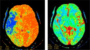
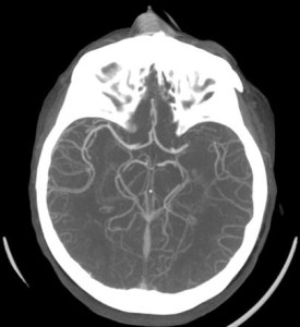
Both CT and MRI studies are essential techniques in the diagnosis and management of patients with stroke.
New techniques and sequences are in turn necessary to establish a more precise diagnosis of patients with stroke. Both vascular radiological tests (CT angiography and MR angiography) as perfusion techniques also called multiparameter (perfusion CT and MRI) are necessary to implement complex treatment of acute and thrombectomy or endovascular techniques.
Allow both demonstrate the level of vascular occlusion and if it still remain viable tissue enough to provide some possible benefit when applying reperfusion therapy.
In most hospitals where intervention is done is the CTA / CT perfusion most used by the faster availability, no incompatibility with pacemakers or other implantable devices or some metal prosthesis.
Besides the CTA is useful in situations where hemorrhagic stroke is important to ensure the absence of injury or underlying malformation responsible for the bleeding (e.g MAV or aneurysm), or even attempt to quantify the risk of rebleeding as ominous signs identifying as the spot-sign.
Also techniques such as CT perfusion allows in cases of absence of risk of vascular occlusion and ischemia such as vasospasm in subarachnoid hemorrhage (SAH) to establish an early diagnosis and initiate treatment to prevent dangerous results of non-treated vasospasm. New software as RAPID (iSchemaView, PA, CA) can get a volume of damage and hipoperfusion with independence techniques and CT machine or brand of software.

