Tag Archives: featured

RACE SCALE / RACECAT

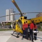
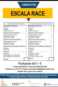
RACE scale (Rapid Artery oClusion Evaluation) try to  detect patients with severe clinical neurological involvement. It is a prehospital and paramedic scale, previous to ER arrival.

A value ≥5 associated with larger tissue impairment and ischemia or hemorrhage risk. It could be associated  to vascular occlusion of large vessel in case it was an ischemic stroke. It could be used in ambulance by paramedical care. But like other scales as pre / Cincinnati or LAMS (Los Angeles) used by paramedics not determine if it is an ischemic or hemorrhagic stroke.

race

    It is important prior training for paramedics to score with this scale, certain values as agnosia, aphasia or bilateral motor impairment can vary  sensitively final score. Also if patient have a previous damage in limb could be scored despite is not due to acute stroke.

The usefulness of these scales (as LAMS, PRE/CINCINNATI, ETC) and target of high specificity and sensitivity are required due to determine which stroke will be candidates for intervention in the future to be referred to high-tech centers or tertiary or comprehensive stroke centers where thrombectomy could be used.

New trial, RACECAT could be started in next monts in Catalonian population to evaluated two possible option drip&ship vs mothership to transfer patients from their home to community hospital supported by telestroke vs directly transfer to a comprehensive stroke center.

ASPECT SCALE

CT is the radiological test of choice at emergency in stroke patients with known onset time. The ability to differentiate incipient lesions on CT emergency reaches 70%. However these injuries should be quantified.

    ASPECTS scale allows a quantitative, way, give a numerical value to a qualitative assessment we value incipient signs of ischemic cerebral involvement.

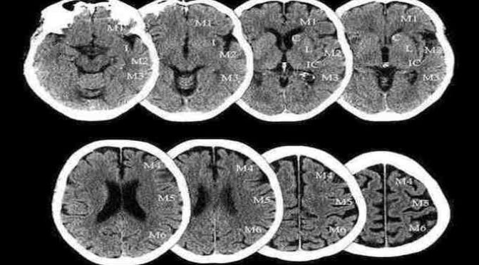
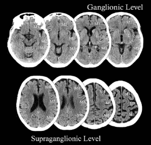
    It is to “assess the lack of differentiation” structures that we usually see in a TAC.  We divide the cerebral hemisphere that has caused damage in 10 segments. (In these segments nuclei of gray and white matter are identified).

    It is evaluated in two slices of the TAC, one just at the level of frontal horns of the lateral ventricle and the other cut in convexity of lateral ventricle. A score of 10 is that we could differentiate all structures. The score  could reach  0 if we identify ten hypodense areas suggestive of infarct as already established. Seven is the cutoff, from> = 7 the patient may be a candidate for endovascular treatment or thrombolysis (equivalent to less 1/3  area of  MCA infarct). Within the same segment is valued worst.

    One way to differentiate structures is lower greey window  to 45HU (Hounsfield units) This window can be more easy and objectively distinguish grey structures and recent infarct as hypodense areas.

aspects

ASPECT score. They are mainly two cuts which to assess the ASPECT score in basal ganglia and above in lateral ventricles but on this tour any hypodense within these territories should be penalized in the final score.

In spite of MR seems more sensitivity to find acute lesions in acute stroke but it requires so much time and patient very relaxed to perform it, this is a reason that get CT a good and reliable test in ER department to evaluate a patient with diagnosis of possible stroke.

However MRI could be a good test in posterior stroke (vertebrobasilar stroke) in acute phase to check it acute lesions. New ingenious scales and score has been mention it to evaluate ischemic lesions in vertebrobasilar portion.

Check other windows of this web www.hubstroke.com to read more about stroke.

NEUROINTERVENTIONISM

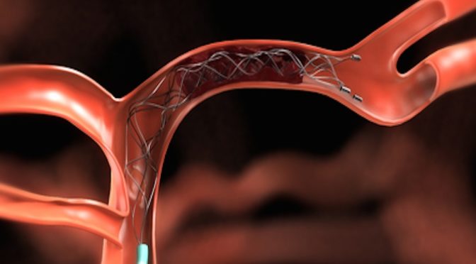
In this section you can check all news and udpate of main studies thatn demonstrated useful of thrombectomy to treat acute stroke.
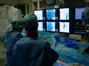
SALA DIVAS

THROMBECTOMY EXCLUSION CRITERIA:

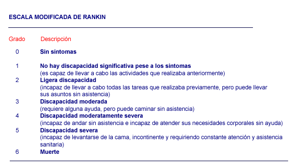
– Rankin in baseline> 2 (the Rankin 3 could be accepted if this limitation is due to osteoarticular disease, neither comorbidity nor cognitive impairment)

– Patient> 85 years. (Between 80-85 whenever this small infarct or early involvement in neuroimaging, baseline Rankin 0-1 and few comorbidity).

– NIH 30 should be prioritized  evaluation of neuroimaging test due to the sedation or intubation.

– More than 8 hours duration since onset time, and is already established infarction without viable tissue present in multimodal techniques (CT or MRI perfusion perfusion). EVEN WITH SMALL CORE INFARCT is recommended thrombectomy enrolled in clinic trials because there is no solid evidence of efficacy in patients over 8 hours of evolution (IIB. NEW)

– Platelets less than 30,000

– Time coagulation INR> 3 or APTT> 2.5 and it is impossible to correct.

– Recent Stroke with size of more than 1/3 MCA territory (ASPECTscore <7) or massive vertebrobasilar (full involvement in neuroimaging section of stem at a level of 50% or more at different levels (mesencephalon-protuberance-bulb). In case of doubt the performance of multiparametric imaging. (MRI, CT perfusion) enable evaluate infarct penumbra areas.  NEW. LOWER LIMIT ASPECTS IT WILL BE 6 (ASPECTS SCORE 0-5 IS NOT RECOMMENDED)

– Bleeding in the hemisphere or in the territory of the same vessel to be treated. (A remote hemorrhagic involvement could not be a contraindication if presented clinical and radiological stability).

– Distal vessel occlusion taht prevent catheter accessibility. NEW RECOMMENDATION 2016: M2 is not recommended because there is no proof, however, in some of the randomized studies included some of these vessels, so most groups agree to treat NIH M2 with high (> 8-10). 

– Severe hemodynamic instability, organ failure or other criteria which the physician considers that the risk outweighs the benefit of this treatment.

– NEW RECOMMENDATION: It is recommended to use devices other than Solitaire or stent-Trevo Retriever. However it may occasionally use different type systems Penumbra (latest models) or situation given anatomy. (Devices like MERCI or Penumbra is not recommended because of its low percentage of recanalization in different studies IIB)
RAPID iSchemaV

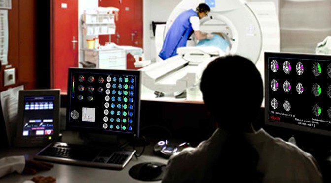
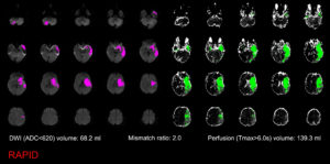
New software or applications are developed to improve feasibility of  CTperfusion with independence  CT machine or individual correction after acquisition.  RAPID is a novedous system of analysis of CT perfusion that provides you exact volume of damage (core infarct) and mismatch (penumbra). It can help to standardise the method to figure out infarct core (irreversible damage) and according to this data and other as age of pacient, NIH score and segment of vessel occlusión evaluated to thrombectomy

iSchemaViem are working in RAPID to improve datas of  CT perfusion. This software is based on several experienced centres treating stroke and many cases of  stroke worldwide. Click and link here RAPID.

3_mr-rapid-output-for-dr-cardona

Check more information in upper Menu bar.